Molecular Cloning and Characterization of E2f3b in Pig
Molecular Cloning and Characterization of E2f3b in Pig
Wentao Wang1,2, Xu Lin3, Jianshu Zhuo3, Dongjie Zhang2, Xiuqin Yang3* and Di Liu1, 2*
Electrophoresis pattern of RT-PCR products. Marker was DL 2000 composed of 2000-, 1000-, 750-, 500-, 250-, and 100-bp DNA fragments.
cDNA sequences of transcript variant 1 of E2F3b in pig. Putative nuclear localization sequences were indicated in bold, and nuclear export signal was boxed. E2F_TDP domain was underlined. E2F_DD domain was shadowed.
Structure of eight transcript variants of porcine E2F3b gene. Boxes indicate exons with closed gray and yellow boxes indicating the sequence added or deleted, respectively. Lines indicate introns. Short vertical lines under boxes indicate the position of start and stop codon sequentially.
Identification of alternative splicing of porcine E2F3b gene by minigene construct. A, Schematic of the minigene construct. Boxes indicate exons, lines indicate introns. Arrows indicate the location of primers. B, Sequence composition of novel alternative splicing variants (NV1-4). The black and yellow boxes indicate the sequences are added and deleted, respectively.
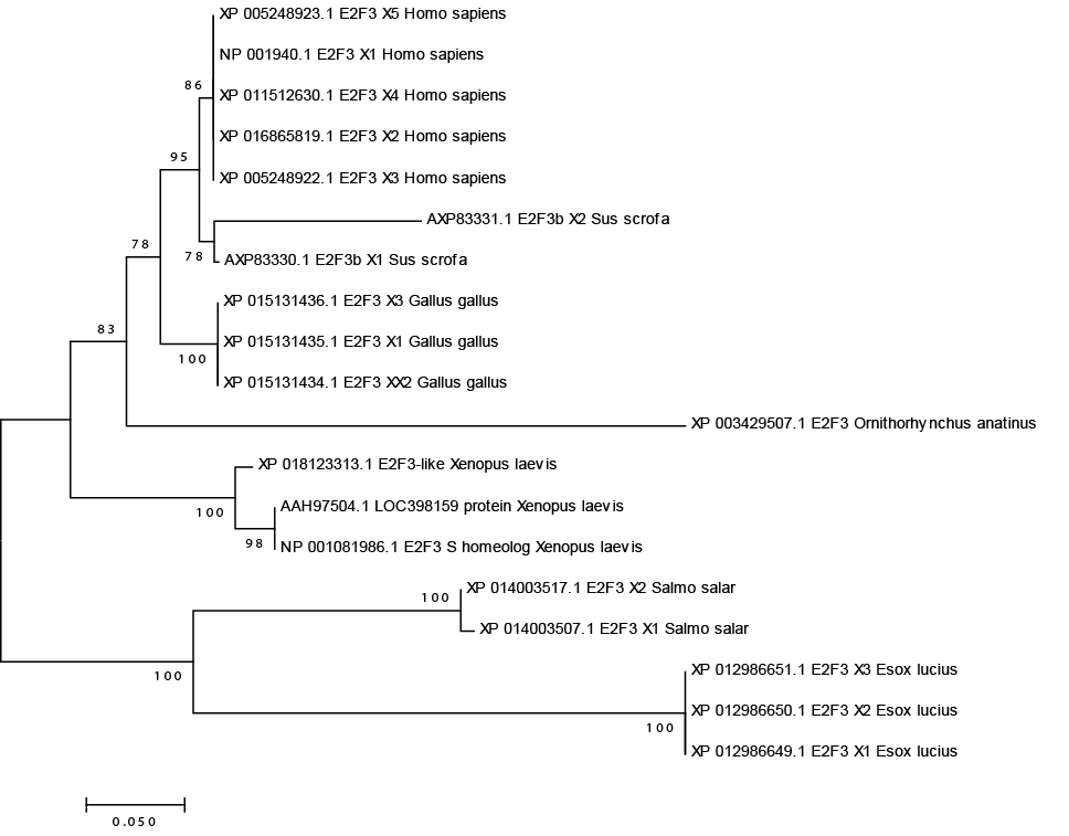
Evolutionary relationships of E2F3 isforms.
Subcellular localization and putative NLSs/NES identification. A, Subcellular localization of two isoforms of porcine E2F3b. B. Effect of putative NLSs/NES on E2F3b subcellular localization. Wt, wild type; Mut, mutant; nt, nucleotide; aa, amino acids. The bar is 100 µm.
Expression profiles of porcine E2F3b. In the upper panel, boxes indicate exons, while closed box indicates intron retained. The slashes indicate skipping of exon 5.


