Polyethylene Glycol (Peg) Mediated In Vitro Characterization of Sugarcane (CP-77/400) Calli and Regenerated Plantlets
Polyethylene Glycol (Peg) Mediated In Vitro Characterization of Sugarcane (CP-77/400) Calli and Regenerated Plantlets
Ayesha Gul1, Mohammad Sayyar Khan1*, Mazhar Ullah1 and Iqbal Munir3
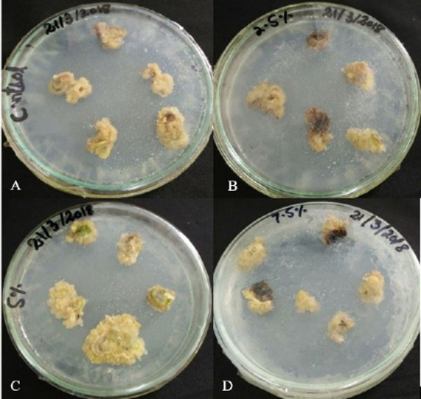
Effect of PEG stress on morphology of sugarcane calli after 30 days of stress application. Calli morphology changed with increasing PEG concentration. Control (0%) (A); 2.5% PEG (B); 5.0% PEG (C); and 7.5% PEG (D).
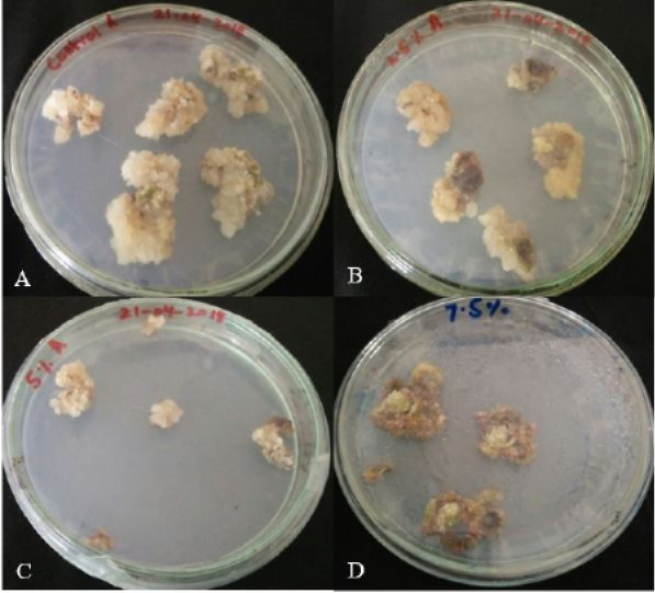
Effect of PEG stress on morphology of sugarcane calli after 60 days of stress application. Calli morphology changed with increasing PEG concentration. Control (0%) (A); 2.5% PEG (B); 5.0% PEG (C); and 7.5% PEG (D).
Regeneration of PEG selected and non-selected sugarcane calli into plantlets. Calli regeneration on MS media (A); growth of plantlets from control calli on media with 0% PEG (B); growth of plantlets from selected calli on media with 7.5% PEG (C), growth of plantlets from non-selected calli on media with 7.5% PEG (D).


