Post-Ischemic Treatment with Sevoflurane: To Reduce Myocardial Ischemic/Reperfusion Injury in Rats by miR-26a-5p/PTEN/PI3K/Akt Signaling Pathway
Post-Ischemic Treatment with Sevoflurane: To Reduce Myocardial Ischemic/Reperfusion Injury in Rats by miR-26a-5p/PTEN/PI3K/Akt Signaling Pathway
Shaolin Zeng, Shuai Yuan* and Kai Liu
Post-ischemic treatment with sevoflurane significantly added miR-26a-5p expression level in rat myocardial tissue. A: Clustering heatmap for post-ischemic treatment with sevoflurane on miRNA in myocardium of rats (n=3). B: The RT-qPCR verified the difference in miRNA expression profile in myocardial tissue of rats after post-ischemic treatment with sevoflurane. C: Venn diagram showed the intersection of miR-26a-5p potential targets in databases of Targetscan, microT-CDS, miRtargetLink and miRpathDB.
PTEN as a direct miR-26a-5p target. A: Effect of post-ischemic treatment with sevoflurane on PTEN mRNA expression (n=3). B (left): Effect of post-ischemic treatment with sevoflurane on the level of PTEN protein (n=3). C (right): The complementary relationship between miR-26a-5p and the 3’UTR of Wild-type PTEN. D: The luciferin reporter groups verified whether miR-26a-5p is a direct PTEN target (n=3). ** represents sham group V.S. I/R model group; ## represents I/R model group V.S. sevoflurane group P<0.01; and represents sevoflurane group V.S. sevoflurane+antamiR-26a-5p group P<0.01.
Post-ischemic treatment with sevoflurane may inhibit PTEN by up-regulating miR-26a-5p to activate the PI3K/Akt pathway. The effect of post-ischemic treatment with sevoflurane on PI3K/Akt signaling pathway (n=3) was detected by Western blot. ** represents sham group V.S. I/R model group; ## represents I/R model group V.S. sevoflurane group P<0.01; and represents sevoflurane group V.S. sevoflurane+antamiR-26a-5p group P<0.01.
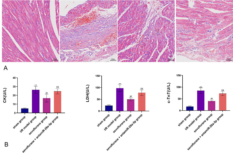
miR-26a-5p inhibition expression level inhibits the myocardial protective effect of sevoflurane after ischemia. A: The effect of miR-26a-5p inhibition expression level on rat myocardial tissue morphology in the sevoflurane group (n = 3). B: The impact of miR-26a-5p inhibition expression level on the content of myocardial tri-enzymes in rat myocardial tissue in the sevoflurane group (n = 7). ** represents sham group V.S. I/R model group; ## represents I/R model group V.S. sevoflurane group P<0.01; and represents sevoflurane group V.S. sevoflurane+antamiR-26a-5p group P<0.01.
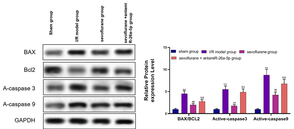
MiR-26a-5p inhibition expression level inhibits the anti-apoptotic effect induced by post-ischemic treatment with sevoflurane. Western blotting was used to examine the impact of inhibition expression of miR-26a-5p on anti-apoptotic effect induced by post-ischemic treatment with sevoflurane (n = 3). ** represents sham group V.S. I/R model group; ## represents I/R model group V.S. sevoflurane group P<0.01; and represents sevoflurane group V.S. sevoflurane+antamiR-26a-5p group P<0.01.
MiR-26a-5p inhibition expression level inhibits the anti-oxidative stress effect of post-ischemic treatment with sevoflurane. A: Influence of antamiR-26a-5p+sevoflurane on the SOD content in myocardial tissue of rats (n = 7). B: Influence of antamiR-26a-5p + sevoflurane on CAT content in rat myocardial tissue (n = 7). C: Influence of antamiR-26a-5p+sevoflurane on GSH-Px content in rat myocardial tissue (n = 7). D: Influence of antamiR-26a-5p + sevoflurane on MDA content in rat myocardial tissue (n = 7). E: Influence of antamiR-26a-5p+sevoflurane on ROS content in rat myocardial tissue (n = 7). ** represents sham group V.S. I/R model group; ## represents I/R model group V.S. sevoflurane group P<0.01; and represents sevoflurane group V.S. sevoflurane+antamiR-26a-5p group P<0.01.
MiR-26a-5p inhibition expression level inhibits the anti-inflammatory effect of post-ischemia treatment with sevoflurane. A: Impact of antamiR-26a-5p+sevoflurane on TNF-α mRNA level in myocardial tissue of rats (n = 7). B: Impact of antamiR-26a-5p+sevoflurane on IL-1β mRNA level in myocardial tissue of rats (N = 7). C: Impact of antamiR-26a-5p+sevoflurane on IL-8 mRNA level in rat myocardial tissue (n = 7). D: Impact of antamiR-26a-5p+sevoflurane on ICAM-1 mRNA level in rat myocardial tissue (n = 7). E: Impact of antamiR-26a-5p+sevoflurane on VCAM-1 mRNA level in rat myocardial tissue (n = 7). We adopted RT-qPCR to detect the influences of different treatment methods on TNF-α, IL-1β, IL-8, ICAM-1 and VCAM-1 mRNA levels in rat myocardial tissue (n = 7). ** represents sham group V.S. I/R model group; ## represents I/R model group V.S. sevoflurane group P<0.01; and represents sevoflurane group V.S. sevoflurane+antamiR-26a-5p group P<0.01.


