

CDX1 Gene Expression in Recurrent Spontaneous Abortion under Extracellular Regulatory Protein Kinase1/2 Signaling Pathway


CDX1 Gene Expression in Recurrent Spontaneous Abortion under Extracellular Regulatory Protein Kinase1/2 Signaling Pathway
Shouyan Cao1, Aili Yan2, Wenhua Zhang3, Fangfang Li1 and Xiaoning Liu4*
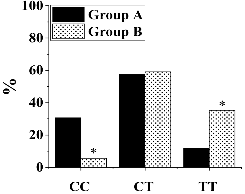
Fig. 1.
Frequency of CDX1 genotypes.
*shows there is a statistically substantial differencecompared with group A (P<0.05). Group A: Non-pcegnant healting female employees (control); Group B: Non-pregnant female patients.
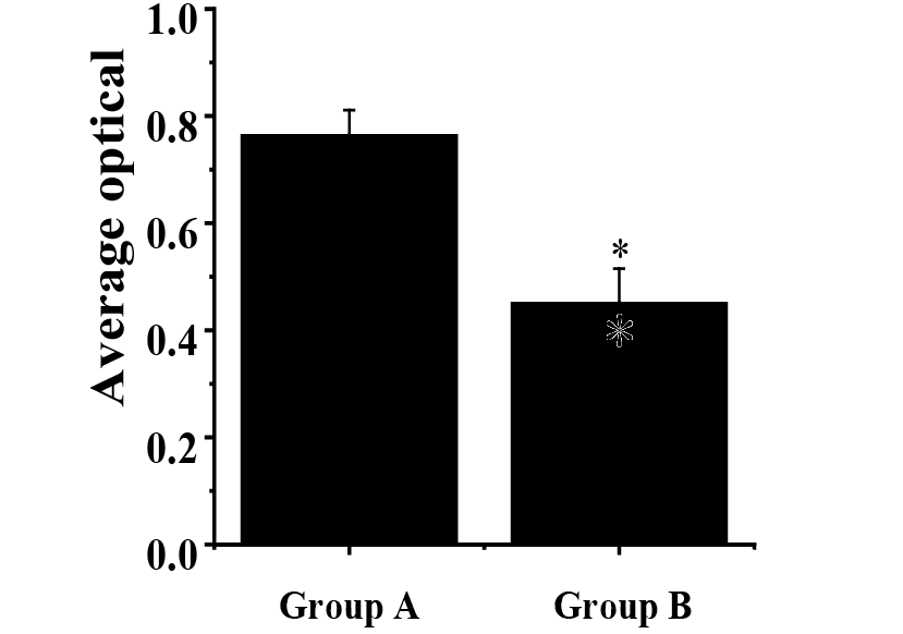
Fig. 2.
Phosphorylation levels of ERK1/2 in subject from both groups.
* shows there is a statistically huge difference compared with group A (P<0.05).
See Figure 1 for details of groups.
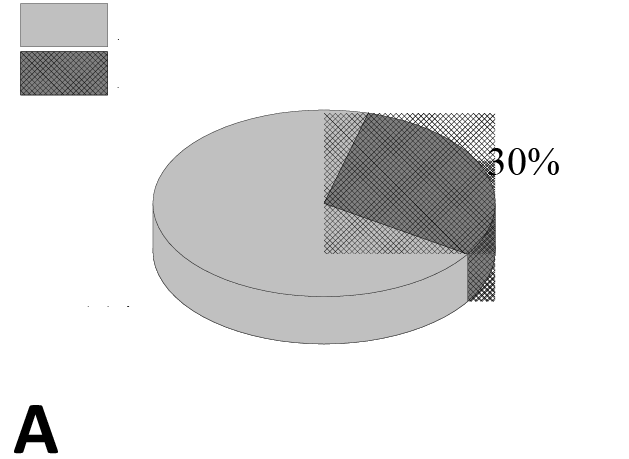
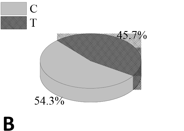
Fig. 3.
CDX1 allele frequency in patients of group A (A), and group B (B).
See Figure 1 for details of groups.
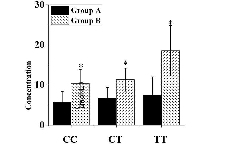
Fig. 4.
Homocysteine and folic acid levels in subjects with the three genotypes.
* shows there is statistically marked differences in contrast to group A (P<0.05).
See Figure 1 for details of groups.
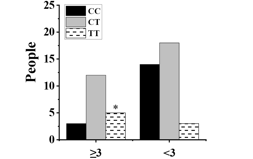
Fig. 6.
Comparison on abortion frequency of different genotypes.
* shows that the difference is statistically considerable in contrast to TT genotype in patients with less than 3 abortions (P<0.05).
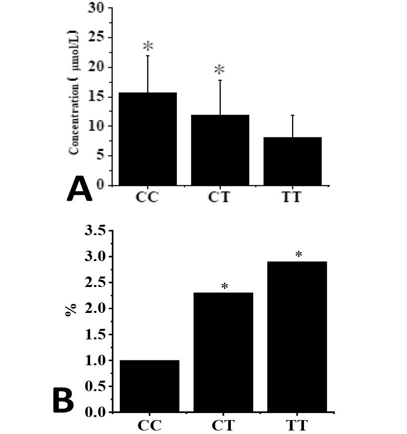
Fig. 5.
Folic acid (A) and RR (B) of three genotypes in group B.
* shows there are statistically great differences in contrast to TT (A) and CC (B) genotype (P<0.05).
See Figure 1 for details of groups.

November 2024
Pakistan J. Zool., Vol. 56
