Submit or Track your Manuscript LOG-IN

Dermatophytosis in Clinically Infected Cats: Diagnoses and Efficacy Therapy

Dermatophytosis in Clinically Infected Cats: Diagnoses and Efficacy Therapy

Alsi Dara Paryuni1, Soedarmanto Indarjulianto2, Tri Untari3 and Sitarina Widyarini4*

1Doctoral Student of Veterinary Science, Faculty of Veterinary Medicine, Universitas Gadjah Mada, Yogyakarta 55281; 2Department of Internal Medicine, Faculty of Veterinary Medicine, Universitas Gadjah Mada, Yogyakarta, 55281; 3Department of Microbiology, Faculty of Veterinary Medicine, Universitas Gadjah Mada, Yogyakarta, 55281; 4Department of Pathology, Faculty of Veterinary Medicine, Universitas Gadjah Mada, Yogyakarta 55281.

 
*Correspondence | Sitarina Widyarini, Department of Pathology, Faculty of Veterinary Medicine, Universitas Gadjah Mada, Yogyakarta 55281; Email: [email protected]

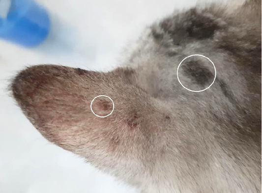
Figure 1:

Lesions from dermatophyte infection in body part of cat with alopecia, erythema, and scale in cat dermatophytosis (white ring).

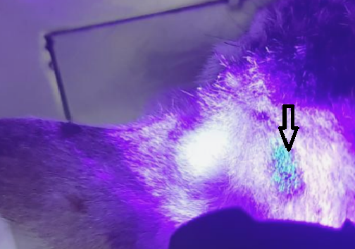
Figure 2:

Ear of cat with fluorescence (apple blue-green color), under Wood’s lamp examination (black arrow).

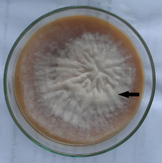
Figure 3:

Fungal colony of Microsporum canis; Cottony, white to buff in colour; with increasing age becomes orange-brown (black arrow).

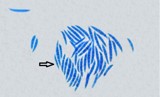
Figure 4:

Microscopic structure of Microsporum canis with Lactophenol cotton blue stain (black arrow).

Figure 5:

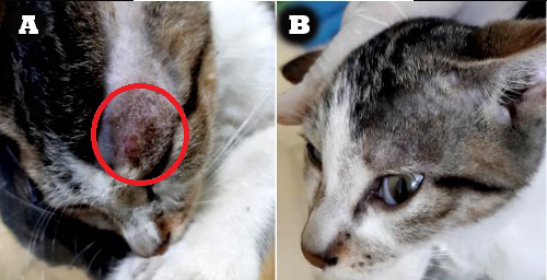
Macroscopic lesions progress (decrease in severity) on treatment with 2% Ketoconazole cream: A. Before treatment (red circle), B. The last day (day 21st) of treatment.

Advances in Animal and Veterinary Sciences

February

Adv. Anim. Vet. Sci., Vol. 13, Iss. 2,

Featuring

Click here for more

Subscribe Today

Receive free updates on new articles, opportunities and benefits


Subscribe Unsubscribe