Developmental Potential of Ovarian Follicles in Mammals: Involvement in Assisted Reproductive Techniques
Developmental Potential of Ovarian Follicles in Mammals: Involvement in Assisted Reproductive Techniques
Abd El-Nasser Ahmed Mohammed*, Aiman Al Mufarji and Sultan Alawaid
Development of primordial follicles during fetal and cycling periods in human.
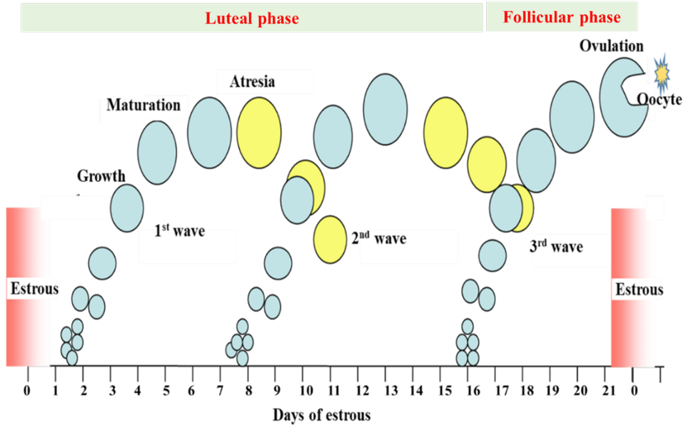
Ovarian cyclic waves during estrous cycle in cattle.
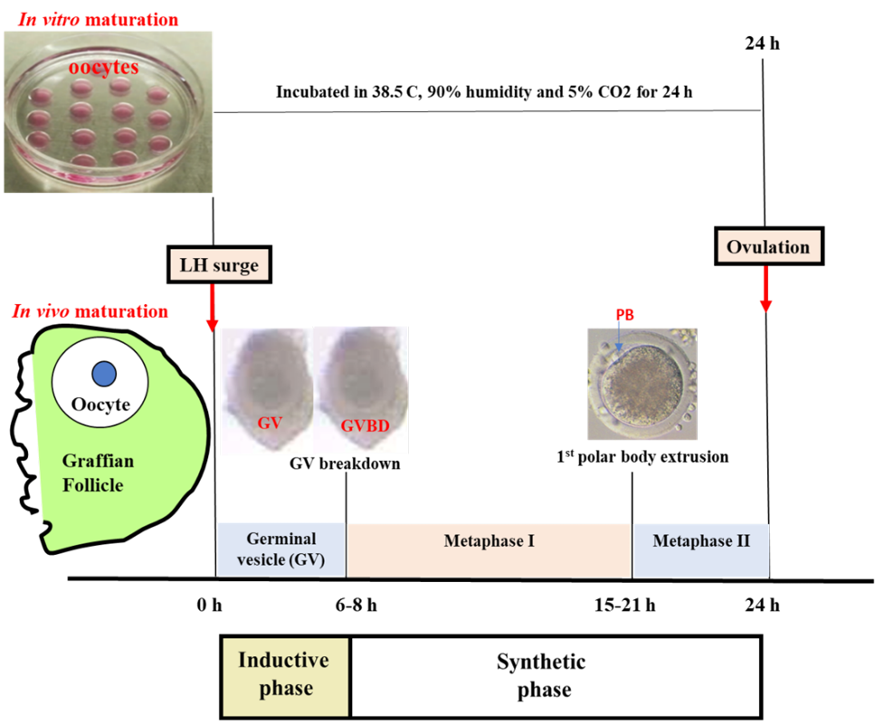
Chronology of events during the maturation of bovine oocytes.
Application of assisted reproductive techniques during fetal, prepubertal and post pubertal periods.
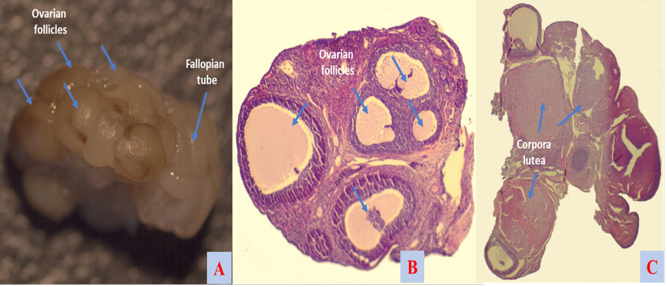
Ovarian morphology and histology upon ovarian transplantation in rats. A, morphology of active transplanted ovary; B, antral follicles of active transplanted ovary; C, corpora lutea of active transplanted ovary.
Maturation and manipulation of mouse oocytes and the resulting outcomes: cumulus-enclosed germinal vesicle (GV) oocytes (A), denuded GV oocytes (B), matured oocytes (C), complete enucleation of germinal vesicle oocyte (D), complete enucleation of cumulus-enclosed germinal vesicle oocyte (E), selective enucleation of g germinal vesicle oocyte (F), enucleation of pro-metaphase I oocyte (G), enucleation of metaphase I oocyte (H), enucleation of metaphase II oocyte (I) enucleolation of germinal vesicle oocyte (J), fetal fibroblast (K), germinal vesicle karyoplasts (L) GV karyoplasts placed under the zona pellucida of enucleated GV oocytes (M) fused with cytoplasts (N, O) developed zygote upon embryonic nuclear transfer (P), developed hatching blastocyst upon embryonic nuclear transfer (Q) offspring upon fallopian embryo transfer.
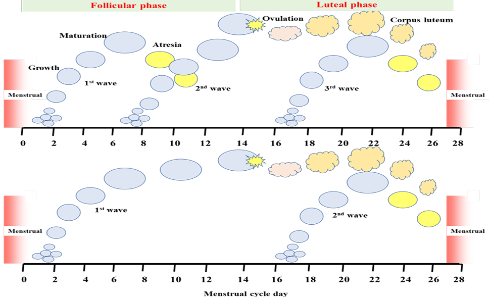
Ovarian cyclic waves during menstrual cycle in human.


