

Distribution and Morphology of Ghrelin-Immunopositive Cells in the Lung of the African Ostrich


Distribution and Morphology of Ghrelin-Immunopositive Cells in the Lung of the African Ostrich
Xiaoting Zhang1,2, Jiaxiang Wang1,2,*, Peng Li1,2,* and Lixun Ye1,2
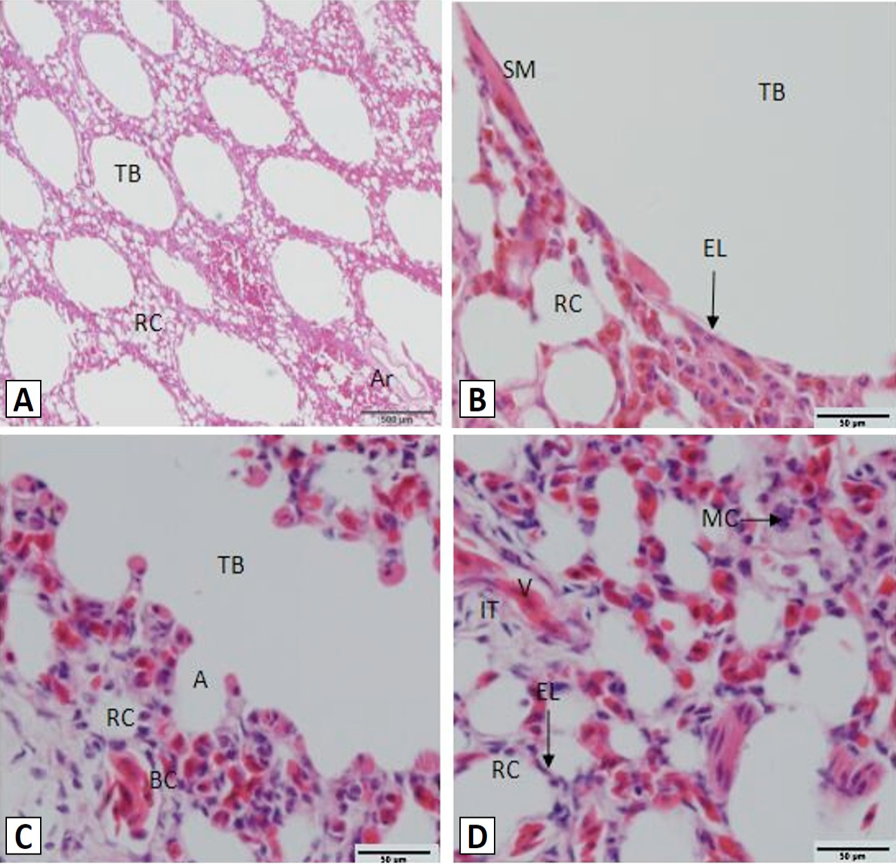
Histology of the African ostrich lungs. A, the lung parenchyma and artery; B, the tertiary bronchus; C, atria; D, respiratory capillaries. TB, tertiary bronchus; RC, respiratory capillaries; Ar, artery; SM, smooth muscle; EL, epithelial layer; A, atria; BC, blood capillary; MC, macrophage; V, vein. Scale bar: A=500 μm; B, C and D=50 μm, Stain: haematoxylin and eosin.

Distribution of ghrelin-ip cells in the lungs of the African ostrich. A, ghrelin-ip cells (arrows) were found within the lung; B, ghrelin-ip cells (arrows) within the tertiary bronchus mucosal epithelial cells; C, ghrelin-ip cells within the respiratory capillary epithelial cells (↑) and macrophage (↑↑); D, the control sections were negative. TB, tertiary bronchus; RC, respiratory capillary; IT, interstitial tissue. Scale bar: A and D=100 μm; B and C= 50 μm.

