Effect of Honey in Improving Breast Cancer Treatment and Gene Expression Modulation of MMPs and TIMPs in Triple-Negative Breast Cancer Cells
Effect of Honey in Improving Breast Cancer Treatment and Gene Expression Modulation of MMPs and TIMPs in Triple-Negative Breast Cancer Cells
Rafa Almeer1,*, A. Alqarni1,2, S. Alqattan3, S. Abdi4,*, S. Alarifi1, Z.Hassan5 and A. Semlali6
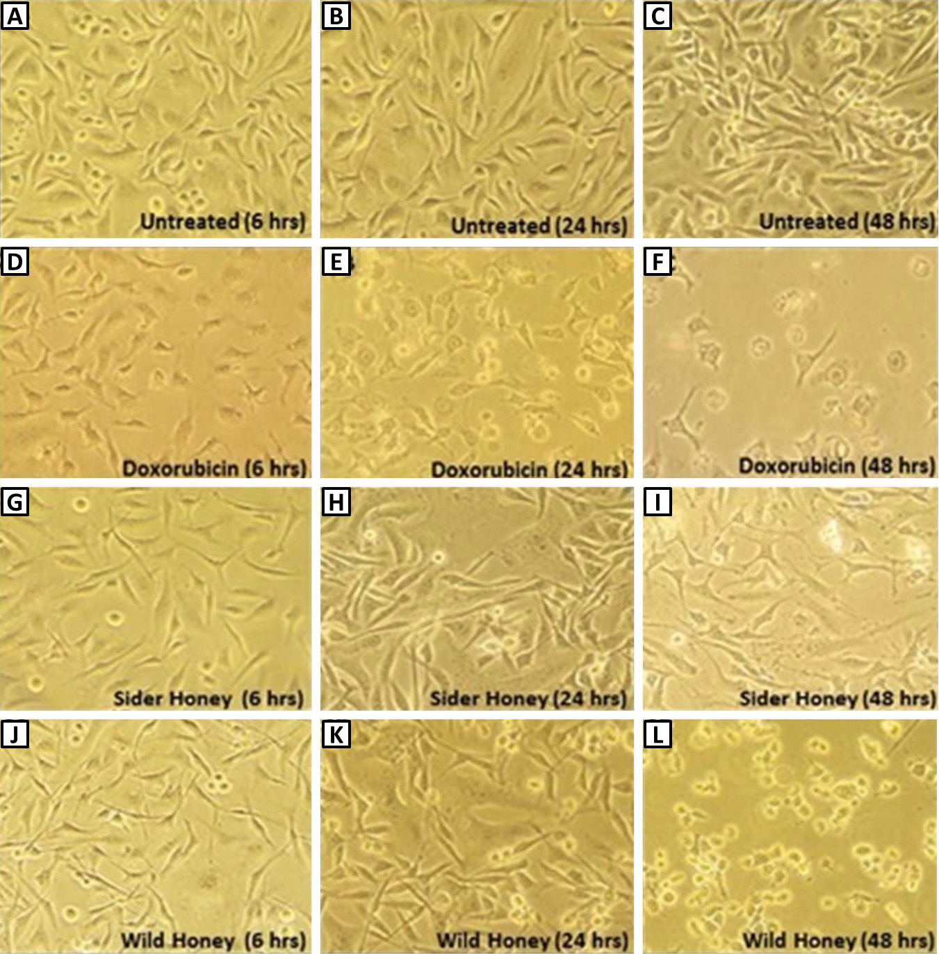
Cell morphology at 6, 24, and 48 h of untreated cells (A-C), post-treatment with doxorubicin 4% (v/v) (D-F), sidr honey 1% (v/v) (G-I) and wild honey 1% (v/v) (J-L).
Cell viability in different subgroups (6, 24, and 48 h) as estimated by MTT assay. The proliferation of MDA-231 cells was detected by MTT assay after treatment with H1, H2, Doxo and compared to untreated cells. *p-value is significant at < 0.05.
Expression of TIMPs in (H1, H2 and doxo) treated and untreated breast cancer MDA-231 cells. A, TIMP-1; B, TIMP-2; C, TIMP-4. Columns, mean (n = 3); SD. *P ≤ 0.05, significant compared to control (untreated cells).
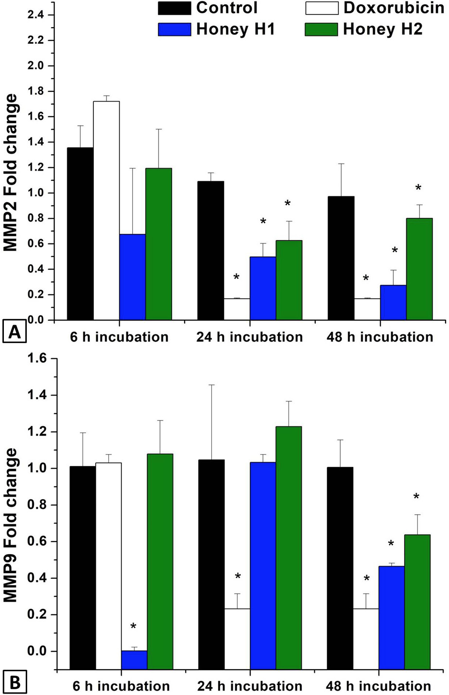
Expression of MMPs in (H1, H2 and Doxo) treated and untreated breast cancer MDA-231 cells. A, MMP-9; B, MMP-2 gene. Columns, mean (n = 3); SD. *p ≤ 0.05, significantly compared to untreated control.


