

Factors Affecting the Absorption of Exosomes by Sertoli Cells


Factors Affecting the Absorption of Exosomes by Sertoli Cells
Ma Hong*, Fu Bo, Wang Liang, Wang Fang, Guo Zhenhua and Liu Di*
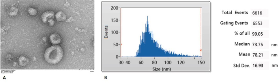
EVs are secreted by STs. (A) Electron microscope image of ST EVs. The scale bar represents 100 nm. (B) Size distribution profile of ST EVs as determined by NTA.
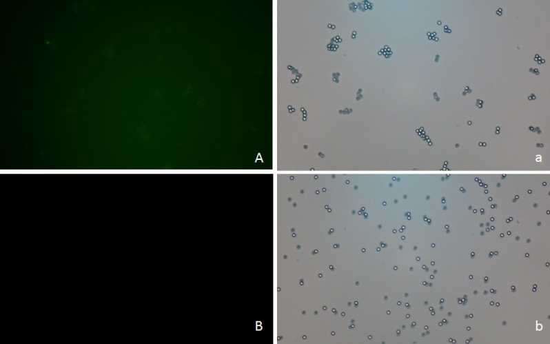
ST-derived EVs were identified by immunofluorescence. The EVs were bound to a 4-μm-diameter latex beads that were in the detection range of the fluorescence microscope. Images were taken under epifluorescence (A, B) and DIC (a, b). (A and a) The EV–bead complexes were bound to fluorescence-conjugated antibody against CD 63. (B) The beads were incubated with fluorescence-conjugated antibody against CD63. (b) Negative control.
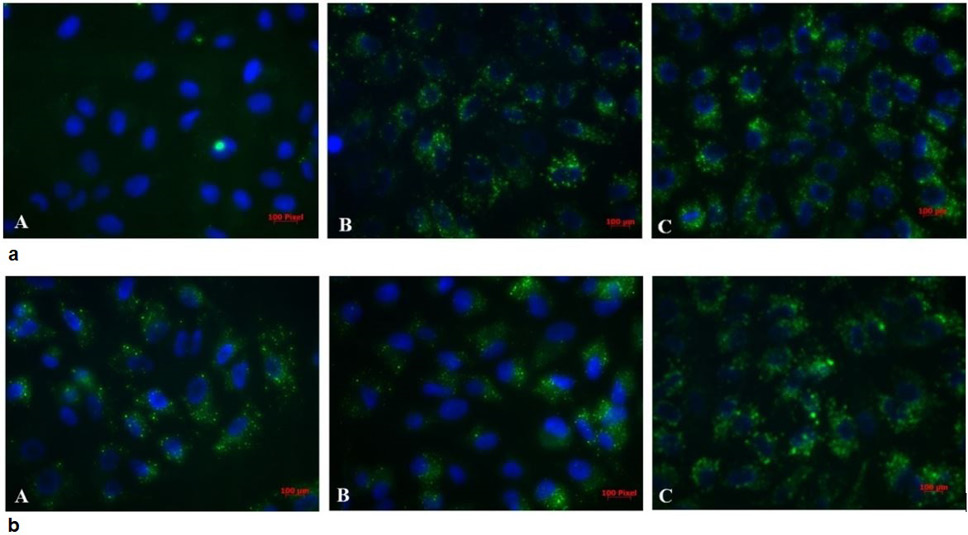
Effect of co-incubation time (a) and temperature (b) the uptake of exosomes by ST cells. (A) DAPI stained nuclei of ST cells; (B) PKH67 stained exosomes; (C) DAPI and PKH67 merged.
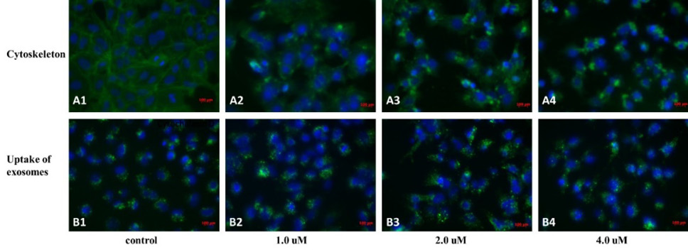
Effect of co-incubation of Cyt D concentrations on PKH67 stained exosomes with ST cells. (A) DAPI stained nuclei of ST cells. (B) PKH67 stained exosomes. (C) DAPI and PKH67 merged.
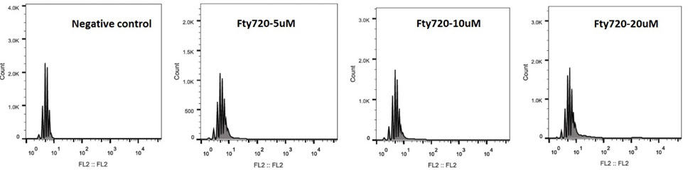
Effects of different concentrations of FTY720 on ST cell apoptosis.
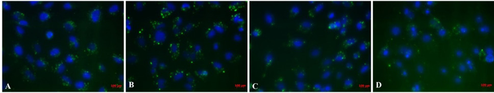
Co-incubation of FTY720 concentrations on PKH67 stained exosomes with ST cells. (A) DAPI stained nuclei of ST cells. (B) PKH67 stained exosomes. (C) DAPI and PKH67 merged.

