

Preparation and Characterization of Iron Oxide Nanoparticles for Effective Epirubicin Delivery to Drug Resistant Breast Cancer Cells


Preparation and Characterization of Iron Oxide Nanoparticles for Effective Epirubicin Delivery to Drug Resistant Breast Cancer Cells
Sayra Tariq, Zeeshan Mutahir and Moazzam Ali*
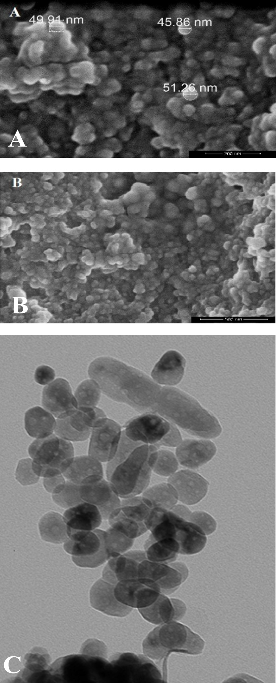
SEM (A, B) and TEM (C) analysis of prepared iron oxide nanoparticles. A, scale bar: 200nm; B, scale bar: 500nm; C, magnification of 80,000X.
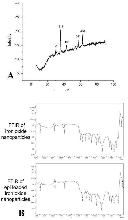
XRD (A) analysis (B) and FTIR spectra of iron oxide nanoparticles, and drug loaded nanoparticles.
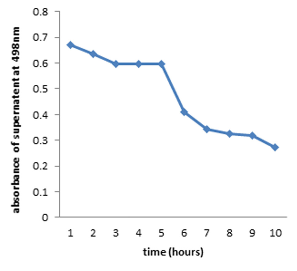
Drug uptake by the particles resulted in reduction of absorbance of drug in the supernatant at 498nm. Particles were suspended in distilled water in an orbital shaker at 37 ℃. 1 ml was removed from the suspension, centrifuged and absorbance of supernatant was taken at 498nm. The mixture was added back to the suspension for further uptake.
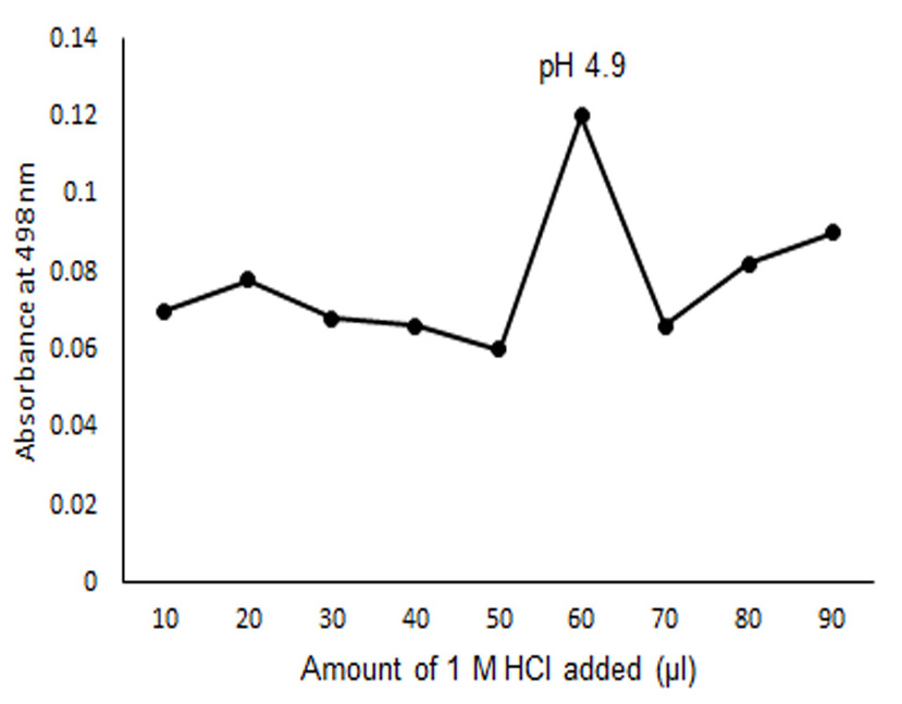
The absorbance of epirubicin in the supernatant at 498nm versus the added amount of 1M HCl in µL (added to 4ml of epirubicin loaded iron oxide nanoparticle suspension in distilled water).
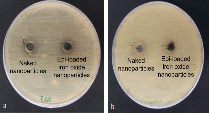
Inhibitory effect of (a) Fe NPs and Epi loaded NPs on E. coli (Gram-negative bacteria) (b) Fe NPs and Epi loaded NPs on S. aureus (Gram positive bacteria) using well diffusion assay. Plates were placed in an incubator for 24 h before measuring the Zones of inhibition by both naked nanoparticles and epirubicin loaded particles.
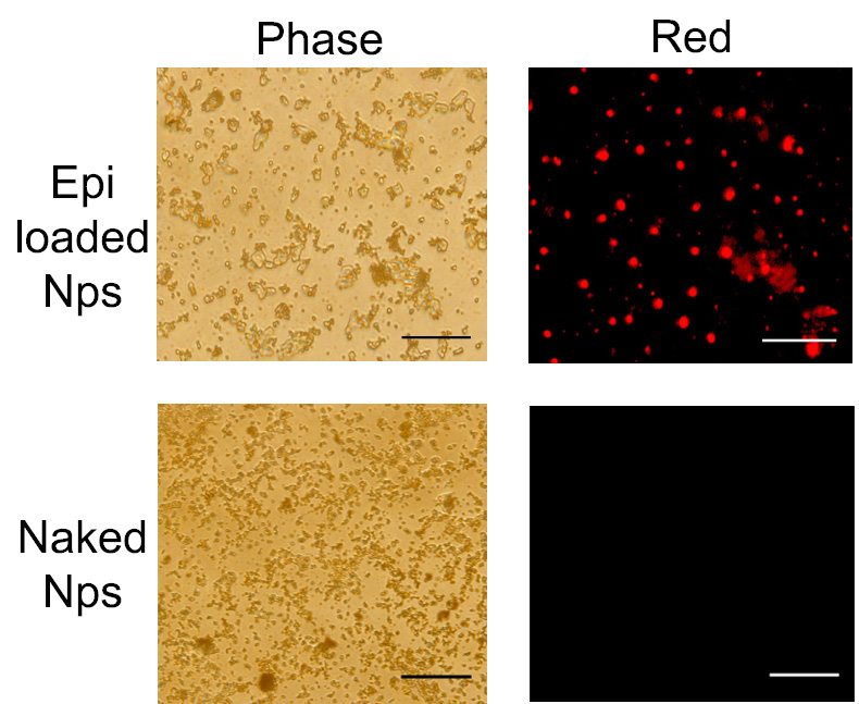
Fluorescent microscopic images of nanoparticles; scale bar 125 µM; nanoparticles (both naked and epirubicin loaded) suspended in distilled water were placed on a slide and were allowed to dry before imaging under the Olympus-BX 51 fluorescence microscope, 40X resolution.
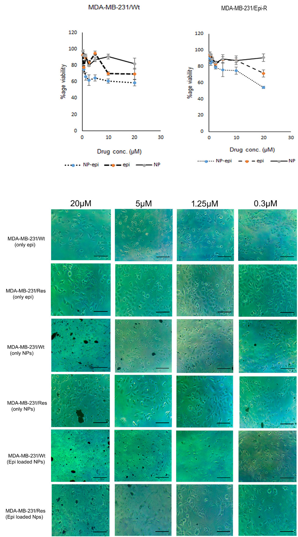
Cell viability of breast cancer cell lines MDA-MB-231/epi R and MDA-MB-231/wt after incubation with epirubicin and epirubicin loaded iron oxide nanoparticles for 72 h, scale bar is 500 µm.
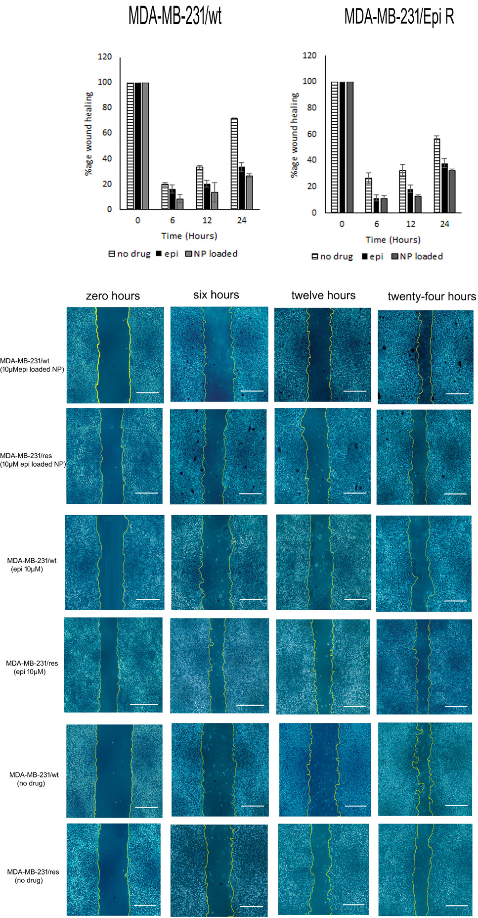
Migration/wound healing assay of MDA-MB-231/wt and MDA-MB-231/Epi resistant cell line. Representative images of wound healing in both cell lines following exposure to 10µM epirubicin alone and epirubicin loaded on iron oxide nanoparticles at zero, six, twelve and twenty-four h. Scale bar 500 µm.

