The Establishment and Evaluation of an Atherosclerotic Vulnerable Plaque Model Involving New Zealand Rabbits
The Establishment and Evaluation of an Atherosclerotic Vulnerable Plaque Model Involving New Zealand Rabbits
Wenxiao Jia1*, Yilinuer Yilihamu1, Yunling Wang1, Shuang Ding1, Dilinuerkezi Aihemaiti1, Hanjiaerbieke Kukun1 and Yanhui Ning2
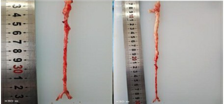
Representative images of the abdominal aorta in the control group and the experimental group. Compared with that in the control group (left panel), the abdominal aorta in the experimental group (right panel) exhibited significant evidence of lipid accumulation and diffuse atherosclerosis.
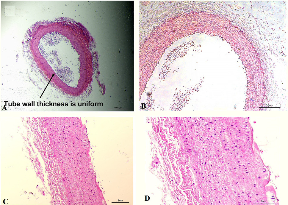
Histological images of the abdominal aorta in the control group: A, abdominal aorta ×10; B, abdominal aorta ×100; C, abdominal aorta ×200; D, abdominal aorta ×400.
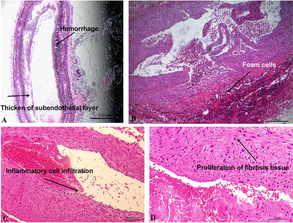
Histological images of the abdominal aorta in the case group: A, abdominal aorta ×10; B, abdominal aorta ×100; C, abdominal aorta ×200; D, abdominal aorta ×400.


