

Ultrastructure of the Tongue of the German Mast Goose (Anser anser) by Scanning Electron Microscopy Before and After Plastination


Ultrastructure of the Tongue of the German Mast Goose (Anser anser) by Scanning Electron Microscopy Before and After Plastination
Saime Betül Baygeldi1, Barıs Can Güzel1*, Ramazan Ilgün2 and Zait Ender Özkan1
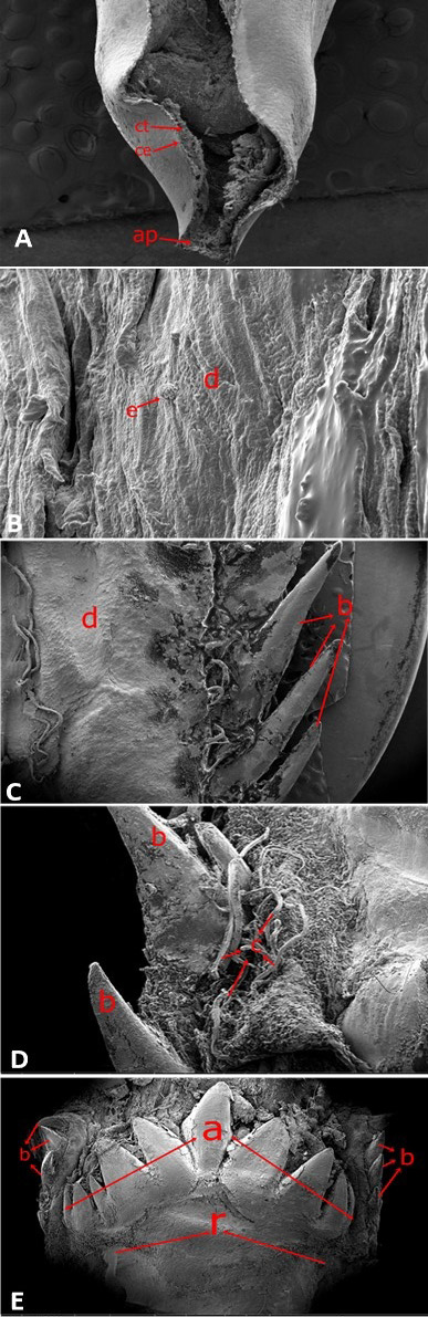
Fig. 1.
Ultrastructure of German Mast excavation tongue. A, cross-sectional SEM view of the apex: ce, Keratimized cpithelium; ap, apex; ct, cartilago. B,C, corpus part: e, para creatimized ridge; d, surface epitholuim, b, papilla linguales; d, surface epitheluim. D, E, radix part: b, papilla linguales caudales; C, papilla filiformes, (a) Papilla conicae, (b) Papilla linguales caudales, (r) Radix.
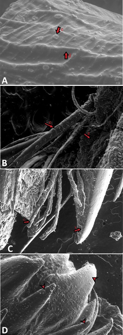
Fig. 2.
Ultrastructure of corpus (A, B) and redix part (C, D) of German Mast excavated tongue (A) and excavation plastine tongue (A, C, D). b, papilla linguales caudales; c, papilla filiformes; d, surface epithelium. Papilla conicae (Arrowhead).
December 2023
Pakistan J. Zool., Vol. 55, Iss. 6, pp. 2501-3000

